Explore
 Validate
 Learn
Learn Western blot
 ELISA
ELISAAntibody data
- Antibody Data
- Antigen structure
- References [0]
- Comments [0]
- Validations
- ELISA [1]
- Immunocytochemistry [1]
Submit
Validation data
Reference
Comment
Report error
- Product number
- ABS 038-15-02 - Provider product page

- Provider
- Invitrogen Antibodies
- Product name
- NGAL Monoclonal Antibody (15)
- Antibody type
- Monoclonal
- Antigen
- Recombinant full-length protein
- Description
- Specificity: ABS 038-15-02 does not cross-react with NGAL from other mammalian species tested. Reactivity: ABS 038-15-02 binds human NGAL in solution and coated onto a solid phase. ABS 038-15-02 can be used as a capture antibody in a sandwich ELISA using biotinylated detection antibodies HYB 211-02-02, ABS 038-04-02, ABS 038-14-02, ABS 038-23-02 and ABS 038-26-02. Using biotinylated ABS 038-15-02 as a detection antibody, the assay reacts equally with NGAL monomer and homodimer on a mass basis. NOTE: Concentration is lot-dependent and can vary from 0.85-1.15 mg/mL
- Reactivity
- Human
- Host
- Mouse
- Isotype
- IgG
- Antibody clone number
- 15
- Vial size
- 400 µL
- Concentration
- 1.02 mg/mL
- Storage
- 4° C, store in dark
No comments: Submit comment
Supportive validation
- Submitted by
- Invitrogen Antibodies (provider)
- Main image
- Experimental details
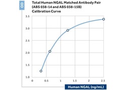
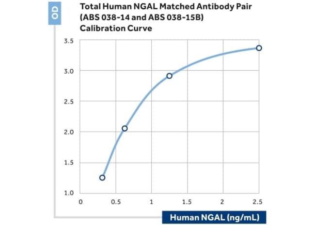
- Sandwich ELISA of NGAL/LCN2 using ABS 038-14 as the capture antibody and ABS 038-15B as the biotinylated detection antibody.
Supportive validation
- Submitted by
- Invitrogen Antibodies (provider)
- Main image
- Experimental details
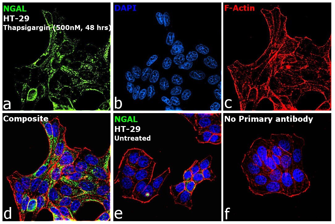
- Immunofluorescence analysis of NGAL was performed using 70% confluent log phase HT-29 treated with Thapsigargin (500 nm, 48hours). The cells were fixed with 4% paraformaldehyde for 10 minutes, permeabilized with 0.1% Triton™ X-100 for 15 minutes, and blocked with 2% BSA for 1 hour at room temperature. The cells were labeled with NGAL Monoclonal Antibody (15) (Product # ABS 038-15-02) at 5 µg/mL in 0.1% BSA, incubated at 4 degree celsius overnight and then labeled with Donkey anti-Mouse IgG (H+L) Highly Cross-Adsorbed Secondary Antibody, Alexa Fluor Plus 488 (Product # A32766, 1/2000), for 45 minutes at room temperature (Panel a: Green). Nuclei (Panel b:Blue) were stained with ProLong™ Diamond Antifade Mountant with DAPI (Product # P36962). F-actin (Panel c: Red) was stained with Rhodamine Phalloidin (Product # R415, 1:300). Panel d represents the merged image showing cytoplasmic localization. Panel e represents untreated cells showing no expression of NGAL Panel f represents control cells with no primary antibody to assess background. The images were captured at 60X magnification.